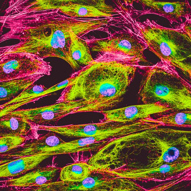
In the realm of cellular biology, accurate assessment of cell viability is crucial for numerous applications, ranging from basic research to clinical diagnostics. One of the key reagents employed in these assessments is 7-Aminoactinomycin D (7-AAD), an Actinomycin D (AD) analog. This vital fluorescent dye has become a staple in flow cytometry due to its ability to distinguish between viable and non-viable cells.
Actinomycin D is the first antibiotic shown to have anti-cancer activity. It serves as an antibiotic and as a chemotherapy medication used to treat a number of types of cancer. It is composed of a natural chemopeptide which is isolated from the bacteria Streptomyces1.
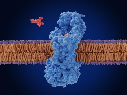
The core utility of 7-AAD , is its action as a fluorescent intercalating agent that binds selectively to GC reach regions of the DNA; thus it is used as a fluorescent marker for DNA in fluorescence microscopy and flow cytometry to differentiate between live and dead cells.
Live cells possess intact cell membranes that exclude 7-AAD, preventing its uptake. In contrast, cells that have undergone apoptosis or necrosis exhibit compromised membranes, allowing 7-AAD to enter and bind to their DNA.
This binding event leads to a strong red fluorescent signal that can be easily detected using a flow cytometer equipped with appropriate filters. When excited by 488 laser light, 7-AAD fluorescence is detected in the far-red range of the spectrum, at 650 nm.
As a potent RNA polymerase inhibitor, 7-AAD was also found to have antibacterial effects. It is impermeable to live cells with intact membranes but can penetrate and bind to the DNA of cells with compromised membranes, typically indicative of late-stage apoptosis or necrosis.
Applications of 7-AAD:
- Cell Viability Assays: One of the primary applications of 7-AAD is in cell viability assays. By staining a cell population with 7-AAD, researchers can quickly and accurately determine the proportion of live versus dead cells. This is particularly useful in experiments where cell death is an important parameter, such as cytotoxicity assays, drug testing, and apoptosis studies.
- Flow Cytometry2-4: In flow cytometry, 7-AAD is frequently used alongside other fluorescent markers to provide a comprehensive analysis of cell populations. For instance, it can be combined with markers for specific cell surface proteins to simultaneously assess cell viability and phenotype.
- Cancer Research: Cancer research often involves assessing the effects of various treatments on tumor cells. 7-AAD staining allows researchers to evaluate the efficacy of anti-cancer drugs by measuring the extent of cell death induced by these treatments. This information is critical for optimizing therapeutic strategies and developing new treatments.
- Clinical Diagnostics: In clinical settings, 7-AAD is used to assess the viability of cells in patient samples. For example, it can be employed in the evaluation of bone marrow or peripheral blood samples to diagnose hematological disorders. Its ability to provide rapid and accurate viability data makes it a valuable tool in diagnostic laboratories.
Advantages of Using 7-AAD:
- High Specificity: 7-AAD high specificity for DNA in cells with compromised membranes ensures accurate discrimination between live and dead cells. This specificity reduces the likelihood of false positives, providing reliable results.
- Ease of Use: The staining protocol for 7-AAD is straightforward and can be easily integrated into existing laboratory workflows. It is compatible with standard flow cytometry procedures, making it accessible to a wide range of researchers.
- Compatibility with Other Stains: 7-AAD can be used in conjunction with other fluorescent dyes and antibodies without significant spectral overlap. This compatibility allows for multiplexing, where multiple parameters can be analyzed simultaneously in a single sample.
While 7-AAD is a powerful tool, there are some considerations and limitations to keep in mind:
- Late Apoptosis and Necrosis Detection: Since 7-AAD primarily stains cells with compromised membranes, it is most effective for detecting late-stage apoptotic and necrotic cells. Early apoptotic cells with intact membranes may not be detected, which could lead to an underestimation of total cell death.
- Photobleaching: Like many fluorescent dyes, 7-AAD is susceptible to photobleaching. Prolonged exposure to light can diminish its fluorescence intensity, potentially affecting the accuracy of results. It is important to minimize light exposure during staining and analysis.
- Optimal Concentration: Determining the optimal concentration of 7-AAD is critical for accurate staining. Excessive concentrations can lead to non-specific binding and increased background fluorescence, while insufficient concentrations may result in weak signals.
An indispensable tool in the field of cell biology, 7-AAD offers a reliable and efficient means of assessing cell viability. Its high specificity, ease of use, and compatibility with other stains make it a preferred choice for researchers and clinicians.
By understanding its mechanism of action and applications, scientists can harness the full potential of 7-AAD to advance research and improve diagnostic capabilities.
For research purposes, Fermentek offers highly pure 7-Aminoactinomycin D (7-AAD), Actinomycin D (>95%) and extra-pure Actinomycin D (>99%) as powder, ready for immediate delivery.
- Finocchiaro G. Actinomycin D: a new opening for an old drug. Neuro Oncol. 2020 Sep 29;22(9):1235-1236. doi: 10.1093/neuonc/noaa172. PMID: 32678904; PMCID: PMC7523453.
- Zembruski NC, Stache V, Haefeli WE, Weiss J. 7-Aminoactinomycin D for apoptosis staining in flow cytometry. Anal Biochem. 2012 Oct 1;429(1):79-81. doi: 10.1016/j.ab.2012.07.005. Epub 2012 Jul 14. PMID: 22796502.
- Wadkins RM, Jovin TM. Actinomycin D and 7-aminoactinomycin D binding to single-stranded DNA. Biochemistry. 1991 Oct 1;30(39):9469-78. doi: 10.1021/bi00103a012. PMID: 1892847.
- Lecoeur H, de Oliveira-Pinto LM, Gougeon ML. Multiparametric flow cytometric analysis of biochemical and functional events associated with apoptosis and oncosis using the 7-aminoactinomycin D assay. J Immunol Methods. 2002 Jul 1;265(1-2):81-96. doi: 10.1016/s0022-1759(02)00072-8. PMID: 12072180.