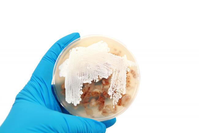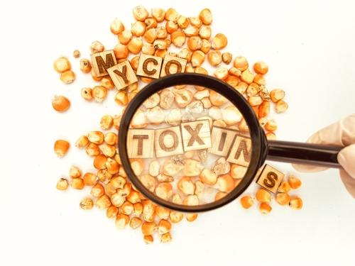
Cytochalasin D is a member of the class of mycotoxins known as Cytochalasins (cytos=cell, chalasis = relaxation), isolated in 19671. Four Cytochalasins were isolated: Cytochalasin A and B from helminthosporium dematioideum, while Cytochalasin C and D from Metarhizium anisopliae1.
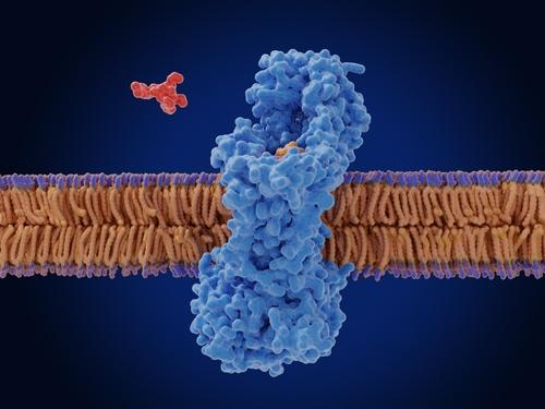
Cytochalasin D is an alkaloid belonging to the macrocyclic lactones family of compounds, with a molecular weight of 507.64 g/mol. It is produced by Helminthosporium and other molds including Aspergillus, Metarhizium, and Zygosporium. Cytochalasin D is a cell-permeable and potent inhibitor of actin polymerization. Actually, it was found to bind to the actin at the filament end, where assembly is taking place, thus inhibiting the rate of actin assembly2. As a result, it disrupts actin polymerization. Thus, Cytochalasin D has become a critical tool in cell biology and pharmacological research.
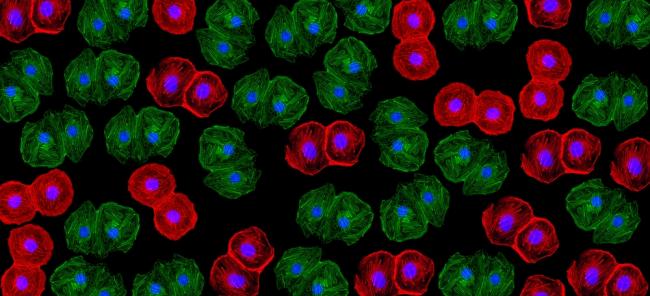
Cytochalasin family of mycotoxins are characterized by their ability to inhibit and influence various cellular processes by targeting actin filaments. Actin is a fundamental component of the cytoskeleton, essential for maintaining cell shape, motility, and division. By binding to the barbed ends of actin filaments, Cytochalasin D prevents further polymerization, leading to significant effects on cellular functions.
Cytochalasin D structure consists of a highly unsaturated 14-membered ring that is crucial for its biological activity. The molecule's ability to bind specifically to actin makes it a valuable tool for studying cellular processes dependent on the actin cytoskeleton.
Applications of Cytochalasin D:
1. Cell Biology Research
Cytochalasin D is extensively used in cell biology to study various aspects of cell structure and function:
- Actin Dynamics: Researchers use Cytochalasin D to investigate the dynamics of actin filaments, including polymerization and .0depolymerization processes. By inhibiting actin polymerization, scientists can observe the resulting changes in cell morphology and motility.
- Cell Motility: The compound is utilized to study cell motility mechanisms, including migration and invasion. By disrupting the actin cytoskeleton, Cytochalasin D helps elucidate the role of actin in these processes.
- Cytokinesis: Cytochalasin D is used to study cytokinesis, the final stage of cell division. By inhibiting actin filament formation, researchers can investigate how cells complete division and the role of the actin cytoskeleton in this process.
2. Pharmacological Research
Cytochalasin D has applications in pharmacological research due to its effects on cellular processes:
- Cancer Research: Cytochalasin D ability to disrupt actin filaments makes it useful in studying cancer cell behavior, particularly cell migration and invasion. Understanding how cancer cells move and invade surrounding tissues is critical for developing anti-metastatic therapies.
- Drug Screening: The compound is used in high-throughput drug screening assays to identify potential inhibitors of actin polymerization. This is particularly relevant in the search for new therapeutic agents targeting the cytoskeleton.
Mechanisms of Action:
1. Actin Polymerization Inhibition
The primary mechanism of action of Cytochalasin D is the inhibition of actin polymerization. It binds to the barbed (plus) ends of actin filaments, preventing the addition of new actin monomers. This leads to the destabilization and eventual depolymerization of existing filaments, resulting in the disruption of the actin cytoskeleton.
2. Disruption of Cellular Processes
By inhibiting actin polymerization, Cytochalasin D affects several cellular processes such as cell shape, structural integrity and morphology; maintaining, cell migration and invasion as well as cell motility. Cytokinesis, the last phase of cell division, is also influenced by Cytochalasin D.
Implications of Cytochalasin D:
1. Research Implications
Cytochalasin D has significantly advanced our understanding of cellular processes involving the actin cytoskeleton. However, its use also has some limitations and concerns:
- Non-Specific Effects: While Cytochalasin D specifically targets actin filaments, it can also have non-specific effects on other cellular components. Researchers must carefully design experiments to account for these potential confounding factors.
- Dose-Dependent Responses: The effects of Cytochalasin D are dose-dependent, with higher concentrations leading to more pronounced disruptions. Accurate dosing is crucial to obtain meaningful and reproducible results.
2. Medical Implications
The potential medical applications of Cytochalasin D are still being explored. While its ability to disrupt actin dynamics holds promise, there are several challenges:
- Toxicity: As a potent mycotoxin, Cytochalasin D can be highly toxic to cells. This limits its use as a therapeutic agent and necessitates the development of derivatives with reduced toxicity.
- Delivery: Effective delivery of Cytochalasin D to target tissues and cells remains a significant challenge. Researchers are investigating various delivery methods to improve its therapeutic potential.
Future Directions and Research:
Development of Derivatives - Ongoing research aims to develop Cytochalasin D derivatives with enhanced specificity and reduced toxicity. These derivatives could provide more effective tools for studying the actin cytoskeleton and potential therapeutic agents for diseases involving actin dynamics.
Exploring New Applications - Researchers are continually exploring new applications for Cytochalasin D in various fields3,4:
- Neuroscience: The actin cytoskeleton plays a crucial role in neuronal development and function. Cytochalasin D can be used to study the role of actin in synaptic plasticity, axon guidance, and neurodegenerative, diseases using stem cells for research.
- Immunology: Actin dynamics are essential for immune cell function. Cytochalasin D can help investigate the role of the cytoskeleton in immune cell migration, activation, and signaling.
- Stem Cells Research in regenerative medicine: Many factors can modulate the differentiation capacity of stem cells, such as Cytochalasins and cyto-permeable mycotoxins, by modifying the actin cellular organization, which can induce a specific and different cell commitment depending on the dose and type of exposed stem cells4.
3. Combination Therapies
Combining Cytochalasin D with other therapeutic agents could enhance its efficacy and reduce toxicity. For example, combining it with drugs targeting other components of the cytoskeleton or signaling pathways could provide synergistic effects, improving therapeutic outcomes.
Cytochalasin D is a powerful tool in cell biology and pharmacological research, providing valuable insights into the role of the actin cytoskeleton in various cellular processes. Its applications in cancer research, drug screening, and understanding cell motility and division have significantly advanced our knowledge. However, the challenges of toxicity and delivery must be addressed to fully realize its potential in medical applications. Ongoing research into derivatives, new applications, and combination therapies holds promise for the future of Cytochalasin D in both research and medicine.
Fermentek offers highly pure Cytochalasin D in various quantities, ready for immediate delivery.
- Aldridge, D.C.; Armstrong, J.J.; Speake, R.N.; Turner, W.B. The Cytochalasins, a new class of biologically active mold metabolites. Chem. Commun. 1967, 1, 26–27.
- Brown, S.S.; Spudich, J.A. Mechanism of action of Cytochalasin: Evidence that it binds to actin filament ends. J. Cell Biol. 1981, 88, 487–491. doi: https://doi.org/10.1083/jcb.88.3.487.
- Sen B, Xie Z, Uzer G, Thompson WR, Styner M, Wu X, Rubin J. Intranuclear Actin Regulates Osteogenesis. Stem Cells. 2015 Oct;33(10):3065-76. doi: 10.1002/stem.2090. PMID: 26140478; PMCID: PMC4788101.
- Pampanella L, Petrocelli G, Abruzzo PM, Zucchini C, Canaider S, Ventura C, Facchin F. Cytochalasins as Modulators of Stem Cell Differentiation. Cells. 2024 Feb 25;13(5):400. doi: 10.3390/cells13050400. PMID: 38474364; PMCID: PMC10931372.